Neural disorders
-
Elevated level of small extracellular vesicules in the serum of patients with depression, epilepsy and epilepsy with depression
Small extracellular vesicles (sEVs) properties and sEVs composition are far from being well-studied for now, especially in the context of mental disorders. To elucidate the role of sEVs in disease we performed a quantitative analysis of the blood sEV in patients with focal epilepsy and patients with focal epilepsy with depression, psychogenic non-epileptic seizures with depression, pure depression, and bipolar affective disorder with the current depressive episode (cDE). Small EVs were isolated from the serum by gel filtration or PEG precipitation, and both methods showed very similar results. Subsequently, we precipitated neuronal sEVs and quantified it with several methods. Activity of lysosomal enzymes was determined in the sEVs fraction. The concentration of the blood sEVs in patients with depression, focal epilepsy, or depression with focal epilepsy was higher than in healthy controls. No difference was found between patients and controls in terms of neuronal sEVs concentration. Another finding of our work is that sEVs in the serum of patients contains various lysosomal enzymes. We suppose that the concentration of the blood sEVs in patients with depression or epilepsy is higher due to the sEVs secretion by the immune cells. Finding sEVs in the blood of patients with depression and focal epilepsy grants validity for future attempts to use sEVs as diagnostic tools for these disorders.
-
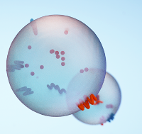
Structural determinants of small extracellular vesicules (exosomes) and their role in biological functions
Extracellular vesicles (EVs) are a new and actively developing area of modern experimental and theoretical biology, which attracts researchers primarily by the possibility of using EVs as diagnostic biomarkers and therapeutic agents. Currently, the greatest amount of data has been accumulated on small extracellular vesicles (sEVs) – exosomes, vesicles of endosomal origin, and ectosomes (previously known as microvesicles), which are the product of direct budding from the plasma membrane. In this review, we address the major steps in the biogenesis of exosomes and ectosomes, the major processes of intracellular membrane trafficking, and signaling involving sEVs. The role of the sEVs in the physiology and pathophysiology of the nervous system is also discussed, as well as many promising aspects of the study of sEVs biology.
-
Quantitative characteristics of small extracellular vesicules from the blood of patients with non-suicidal self-harm
Objective of the study – determination of quantitative characteristics of small extracellular vesicles (sEV) in the blood of patients with non-suicidal self-injury (NSSI) and comparison of the concentration and size of sEV’s in patients with major depressive disorder (MDD) with and without NSSI, as well as an assessment of the relationship between the sizes and concentrations of sEV in the sample with such parameters as the severity of situational and personal anxiety, depression and suicidal risk. The study included 28 patients (11 m./17 f.) with a current episode of major depression and at least five episodes of NSSI in the last 12 months (main group, mean age 28.3 years) and 28 patients with major depression identical in sex and age without NSSI throughout life (comparison group). Patient mental status was assessed using the MINI interview, the Beck Depression Inventory II (BDI II), and the Spielberger Anxiety Scale. Isolation of sEV from blood was carried out using polyethylene glycol (PEG) precipitation and gel filtration. The size and concentration of isolated particles were estimated using dynamic light scattering (DLS) and nanoparticle tracking analysis (ATN). The groups differed significantly in the severity of depression according to the BDI-II questionnaire, the Spielberger Situational Anxiety Scale, and the Spielberger Personality Anxiety Scale. The assessment of suicidal risk, carried out according to the corresponding module of the MINI questionnaire, revealed a significantly larger number of participants with medium and high suicidal risk in the group of patients with NSSI. The sEV fraction was isolated from the blood of the patients of the main group and the comparison group. There were no differences in the concentration and size of sEV between groups of patients with depression with and without NSSI. In our study, the dependence of the concentration and size of sEV on the severity of depression, situational and personal anxiety, and the severity of suicidal risk wasn’t revealed.
Conclusion: NSSI in individuals with major depressive disorder is associated with a more severe course of the disorder (greater severity of depression, situational and personal anxiety), as well as a higher risk of suicide. Our study did not reveal any differences in the quantitative characteristics of sEV in patients with a depressive episode with and without NSSI. Future studies should focus on investigating the structural differences and functional features of sEV in NSSI.
-
Lower loss rate of serotonergically modulated neuronal accumulator for time in patients with major depressive disorder
Major depressive disorder (MDD) is characterized by dramatic and persistent worsening of mood, as well as a subjective feeling of time slowing. However, experimental data on time perception are inconsistent. As serotonergic dysfunction implicated in MDD etiology, we aim to examine time perception in MDD through the framework of lossy temporal integration model, previously also related to serotonergic transmission. Thirty-one patients with recurrent depressive disorder in partial remission and thirty control participants, without a history of psychiatric and neurological disorders, performed duration discrimination of visual stimuli (duration ranges from 3.2 to 6.4 s) and subjective minute production tasks. To infer about central serotonergic transmission, an electroencephalogram in response to the 1000 Hz tone of different intensity (50, 60, 70 and 80 dB SPL) was recorded. Patients with MDD shorten the past durations in the duration discrimination task significantly less than controls, thus being more objective. No difference in the subjective minute production was recorded. Patients with MDD have also exhibited larger auditory evoked potentials in response to the tones of high intensity (70 and 80 dB SPL) when compared with the controls. This resulted in a steeper slope of auditory evoked potentials by intensity function. These converging findings suggest a lower loss rate of neuronal temporal accumulator modulated by serotonergic transmission in patients with MDD.
-
Abnormal spectral and scale-free properties of resting-state EEG in girls with Rett syndrome
Spontaneous EEG contains important information about neuronal network properties that is valuable for understanding different neurological and psychiatric conditions. Rett syndrome (RTT) is a rare neurodevelopmental disorder, caused by mutation in the MECP2 gene. RTT is characterized by severe motor impairments that prevent adequate assessment of cognitive functions. Here we probe EEG parameters obtained in no visual input condition from a 28-channels system in 23 patients with Rett Syndrome and 38 their typically developing peers aged 3–17 years old. Confirming previous results, RTT showed a fronto-central theta power (4–6.25 Hz) increase that correlates with a progression of the disease. Alpha power (6.75–11.75 Hz) across multiple regions was, on the contrary, decreased in RTT, also corresponding to general background slowing reported previously. Among novel results we found an increase in gamma power (31–39.5 Hz) across frontal, central and temporal electrodes, suggesting elevated excitation/inhibition ratio. Long-range temporal correlation measured by detrended fluctuation analysis within 6–13 Hz was also increased, pointing to a more predictable oscillation pattern in RTT. Overall measured EEG parameters allow to differentiate groups with high accuracy, ROC AUC value of 0.92 ± 0.08, indicating clinical relevance.
-
Does the Potocki–Lupski Syndrome Convey the Autism Spectrum Disorder Phenotype? Case Report and Scoping Review
Potocki–Lupski Syndrome (PTLS) is a rare condition associated with a duplication of 17p11.2 that may underlie a wide range of congenital abnormalities and heterogeneous behavioral phenotypes. Along with developmental delay and intellectual disability, autism-specific traits are often reported to be the most common among patients with PTLS. To contribute to the discussion of the role of autism spectrum disorder (ASD) in the PTLS phenotype, we present a case of a female adolescent with a de novo dup(17) (p11.2p11.2) without ASD features, focusing on in-depth clinical, behavioral, and electrophysiological (EEG) evaluations. Among EEG features, we found the atypical peak–slow wave patterns and a unique saw-like sharp wave of 13 Hz that was not previously described in any other patient. The power spectral density of the resting state EEG was typical in our patient with only the values of non-linear EEG dynamics: Hjorth complexity and fractal dimension were drastically attenuated compared with the patient’s neurotypical peers. Here we also summarize results from previously published reports of PTLS that point to the approximately 21% occurrence of ASD in PTLS that might be biased, taking into account methodological limitations. More consistent among PTLS patients were intellectual disability and speech and language disorders.
-
Brain oscillatory patterns of affective prosody perception in children with autism spectrum disorder
Background
Paralinguistic features, such as prosody (tempo, loudness, and timbre), are an essential marker of a speaker’s emotional state. Abnormal processing of emotional prosody may result in the deficient social behavior associated with autism spectrum disorder (ASD).
Method
Two groups of children participated in our study: the ASD group consisted of 30 preschoolers from 4 to 6 years of age and 24 typically developing (TD) peers. An electroencephalogram (EEG) was acquired in response to a combination of syllables uttered with the following types of emotional prosody: joy, anger, sadness, fear, and calmness.
Results
Children with ASD and TD showed a similar EEG oscillatory response to fear and anger prosodies. Significant group differences in power spectral density (PSD) were detected for sad and joy intonations. The PSD differences between pairs of intonations, such as joyful and sad, sad and neutral, or joyful and neutral, were significantly higher in the control group than in the ASD group. EEG responses to affective prosody also demonstrated less hemispheric asymmetry in the ASD than in the TD group.
Conclusions
Our results suggest that difficulties in emotional prosody recognition in autistic children could be based on the atypical processing of specific acoustic features coding differences between sad, neutral, and joyful intonations and could underlie emotional perception deficits in individuals with ASD.
Elevated Serum Cortisol Levels in Patients with Focal Epilepsy, Depression, and Comorbid Epilepsy and Depression
Background: The hypothalamic-pituitary-adrenal (HPA) axis, inflammatory processes and neurotrophic factor systems are involved in pathogenesis of both epilepsy and depressive disorders. The study aimed to explore these systems in patients with focal epilepsy (PWE, n = 76), epilepsy and comorbid depression (PWCED n = 48), and major depressive disorder (PWMDD, n = 62) compared with healthy controls (HC, n = 78). Methods: Parameters of the HPA axis, neurotrophic factors, and TNF-α were measured in blood serum along with the hemogram. Results: Serum cortisol level was augmented in PWE, PWCED, and PWMDD compared with HC and was higher in PWMDD than in PWE. Serum cortisol negatively correlated with Mini–Mental State Examination (MMSE) score in PWE, and positively with depression inventory–II (BDI-II) score in PWMDD. Only PWMDD demonstrated elevated plasma ACTH. Serum TNF-α, lymphocytes, and eosinophils were augmented in PWMDD; monocytes elevated in PWE and PWCED, while neutrophils were reduced in PWE and PWMDD. Serum BDNF was decreased in PWE and PWCED, CNTF was elevated in all groups of patients. In PWE, none of above indices depended on epilepsy etiology. Conclusions: The results confirm the involvement of HPA axis and inflammatory processes in pathogenesis of epilepsy and depression and provide new insights in mechanisms of epilepsy and depression comorbidity.
Brain Trauma, Glucocorticoids and Neuroinflammation: Dangerous Liaisons for the Hippocampus
Glucocorticoid-dependent mechanisms of inflammation-mediated distant hippocampal damage are discussed with a focus on the consequences of traumatic brain injury. The effects of glucocorticoids on specific neuronal populations in the hippocampus depend on their concentration, duration of exposure and cell type. Previous stress and elevated level of glucocorticoids prior to pro-inflammatory impact, as well as long-term though moderate elevation of glucocorticoids, may inflate pro-inflammatory effects. Glucocorticoid-mediated long-lasting neuronal circuit changes in the hippocampus after brain trauma are involved in late post-traumatic pathology development, such as epilepsy, depression and cognitive impairment. Complex and diverse actions of the hypothalamic–pituitary–adrenal axis on neuroinflammation may be essential for late post-traumatic pathology. These mechanisms are applicable to remote hippocampal damage occurring after other types of focal brain damage (stroke, epilepsy) or central nervous system diseases without obvious focal injury. Thus, the liaisons of excessive glucocorticoids/dysfunctional hypothalamic–pituitary–adrenal axis with neuroinflammation, dangerous to the hippocampus, may be crucial to distant hippocampal damage in many brain diseases. Taking into account that the hippocampus controls both the cognitive functions and the emotional state, further research on potential links between glucocorticoid signaling and inflammatory processes in the brain and respective mechanisms is vital.
A Combined Effect of G-Quadruplex and Neuro-Inducers as an Alternative Approach to Human Glioblastoma Therapy
Cancer cell reprogramming based on treatment with G-quadruplex, having antiproliferative power, along with small molecules able to develop iPSCs into neurons, could create a novel approach to diminish the chance of glioblastoma recurrence and circumvent tumor resistance to conventional therapy. In this research, we have tested several combinations of factors to affect both total cell cultures, derived from tumor tissue of patients after surgical resection and two subfractions of this cell culture after dividing them into CD133-enriched and CD133-depleted populations (assuming CD133 to be a marker of glioblastoma stem-like cells). CD133+ and CD133- cells exhibit different responses to the same combinations of factors; CD133+ cells have stem-like properties and are more resistant. Therefore, the ability to affect CD133+ cells provides a possibility to circumvent resistance to conventional therapy and to build a promising strategy for translation to improve the treatment of patients with glioblastoma.
Neuroinflammation and Neuronal Loss in the Hippocampus Are Associated with Immediate Posttraumatic Seizures and Corticosterone Elevation in Rats
Hippocampal damage after traumatic brain injury (TBI) is associated with late posttraumatic conditions, such as depression, cognitive decline and epilepsy. Mechanisms of selective hippocampal damage after TBI are not well understood. In this study, using rat TBI model (lateral fluid percussion cortical injury), we assessed potential association of immediate posttraumatic seizures and changes in corticosterone (CS) levels with neuroinflammation and neuronal cell loss in the hippocampus. Indices of distant hippocampal damage (neurodegeneration and neuroinflammation) were assessed using histological analysis (Nissl staining, Iba-1 immunohistochemical staining) and ELISA (IL-1β and CS) 1, 3, 7 and 14 days after TBI or sham operation in male Wistar rats (n = 146). IL-1β was elevated only in the ipsilateral hippocampus on day 1 after trauma. CS peak was detected on day 3 in blood, the ipsilateral and contralateral hippocampus. Neuronal cell loss in the hippocampus was demonstrated bilaterally; in the ipsilateral hippocampus it started earlier than in the contralateral. Microglial activation was evident in the hippocampus bilaterally on day 7 after TBI. The duration of immediate seizures correlated with CS elevation, levels of IL-1β and neuronal loss in the hippocampus. The data suggest potential association of immediate post-traumatic seizures with CS-dependent neuroinflammation-mediated distant hippocampal damage.
Facial emotion perception in young female students with subsyndromal panic disorder. Behavioral and ERP study
This study aimed to examine the behavioral and neural correlates of facial emotion recognition in young women with subsyndromal panic disorder (SPD). In the experiment 15 non-medicated women with SPD and 17 matched healthy controls were tasked with recognizing angry, fearful, happy, and neutral facial expressions, and accuracy, reaction time (RT), and event-related potentials (ERPs) were recorded. Significant between-group differences in behavioral characteristics (accuracy of emotion recognition and RT) were not found, however, the SPD subjects demonstrated a slower response to fearful expressions compared to neutral and happy expressions. More distinct between-group differences were observed for the EPRs. The SPD subjects demonstrated increased amplitudes of the P100 ERP component in the occipital area and the P200 component over the occipital and temporal regions. In the frontal regions the SPD group showed a greater amplitude of the N200 component and also an increased negativity 350−450 ms after stimulus presentation. According to the dipole source modeling data, the SPD subjects showed enhanced activation in the extra-striate cortex which increased in intensity when angry and fearful faces were presented. Thus, young women with SPD which manifested in infrequent panic attacks showed significant alterations in ERP characteristics of emotional processing, which may be considered as a more sensitive indicator of early-stage panic disorder than the observed behavioral measures.