Molecular biology
-
Elevated level of small extracellular vesicules in the serum of patients with depression, epilepsy and epilepsy with depression
Small extracellular vesicles (sEVs) properties and sEVs composition are far from being well-studied for now, especially in the context of mental disorders. To elucidate the role of sEVs in disease we performed a quantitative analysis of the blood sEV in patients with focal epilepsy and patients with focal epilepsy with depression, psychogenic non-epileptic seizures with depression, pure depression, and bipolar affective disorder with the current depressive episode (cDE). Small EVs were isolated from the serum by gel filtration or PEG precipitation, and both methods showed very similar results. Subsequently, we precipitated neuronal sEVs and quantified it with several methods. Activity of lysosomal enzymes was determined in the sEVs fraction. The concentration of the blood sEVs in patients with depression, focal epilepsy, or depression with focal epilepsy was higher than in healthy controls. No difference was found between patients and controls in terms of neuronal sEVs concentration. Another finding of our work is that sEVs in the serum of patients contains various lysosomal enzymes. We suppose that the concentration of the blood sEVs in patients with depression or epilepsy is higher due to the sEVs secretion by the immune cells. Finding sEVs in the blood of patients with depression and focal epilepsy grants validity for future attempts to use sEVs as diagnostic tools for these disorders.
-
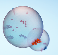
Structural determinants of small extracellular vesicules (exosomes) and their role in biological functions
Extracellular vesicles (EVs) are a new and actively developing area of modern experimental and theoretical biology, which attracts researchers primarily by the possibility of using EVs as diagnostic biomarkers and therapeutic agents. Currently, the greatest amount of data has been accumulated on small extracellular vesicles (sEVs) – exosomes, vesicles of endosomal origin, and ectosomes (previously known as microvesicles), which are the product of direct budding from the plasma membrane. In this review, we address the major steps in the biogenesis of exosomes and ectosomes, the major processes of intracellular membrane trafficking, and signaling involving sEVs. The role of the sEVs in the physiology and pathophysiology of the nervous system is also discussed, as well as many promising aspects of the study of sEVs biology.
-
Quantitative characteristics of small extracellular vesicules from the blood of patients with non-suicidal self-harm
Objective of the study – determination of quantitative characteristics of small extracellular vesicles (sEV) in the blood of patients with non-suicidal self-injury (NSSI) and comparison of the concentration and size of sEV’s in patients with major depressive disorder (MDD) with and without NSSI, as well as an assessment of the relationship between the sizes and concentrations of sEV in the sample with such parameters as the severity of situational and personal anxiety, depression and suicidal risk. The study included 28 patients (11 m./17 f.) with a current episode of major depression and at least five episodes of NSSI in the last 12 months (main group, mean age 28.3 years) and 28 patients with major depression identical in sex and age without NSSI throughout life (comparison group). Patient mental status was assessed using the MINI interview, the Beck Depression Inventory II (BDI II), and the Spielberger Anxiety Scale. Isolation of sEV from blood was carried out using polyethylene glycol (PEG) precipitation and gel filtration. The size and concentration of isolated particles were estimated using dynamic light scattering (DLS) and nanoparticle tracking analysis (ATN). The groups differed significantly in the severity of depression according to the BDI-II questionnaire, the Spielberger Situational Anxiety Scale, and the Spielberger Personality Anxiety Scale. The assessment of suicidal risk, carried out according to the corresponding module of the MINI questionnaire, revealed a significantly larger number of participants with medium and high suicidal risk in the group of patients with NSSI. The sEV fraction was isolated from the blood of the patients of the main group and the comparison group. There were no differences in the concentration and size of sEV between groups of patients with depression with and without NSSI. In our study, the dependence of the concentration and size of sEV on the severity of depression, situational and personal anxiety, and the severity of suicidal risk wasn’t revealed.
Conclusion: NSSI in individuals with major depressive disorder is associated with a more severe course of the disorder (greater severity of depression, situational and personal anxiety), as well as a higher risk of suicide. Our study did not reveal any differences in the quantitative characteristics of sEV in patients with a depressive episode with and without NSSI. Future studies should focus on investigating the structural differences and functional features of sEV in NSSI.
-
Glucocorticoids Orchestrate Adult Hippocampal Plasticity: Growth Points and Translational Aspects
The review analyzes modern concepts about the control of various mechanisms of the hippocampal neuroplasticity in adult mammals and humans by glucocorticoids. Glucocorticoid hormones ensure the coordinated functioning of key components and mechanisms of hippocampal plasticity: neurogenesis, glutamatergic neurotransmission, microglia and astrocytes, systems of neurotrophic factors, neuroinflammation, proteases, metabolic hormones, neurosteroids. Regulatory mechanisms are diverse; along with the direct action of glucocorticoids through their receptors, there are conciliated glucocorticoid-dependent effects, as well as numerous interactions between various systems and components. Despite the fact that many connections in this complex regulatory scheme have not yet been established, the study of the factors and mechanisms considered in the work forms growth points in the field of glucocorticoid-regulated processes in the brain and primarily in the hippocampus. These studies are fundamentally important for the translation into the clinic and the potential treatment/prevention of common diseases of the emotional and cognitive spheres and respective comorbid conditions.
-
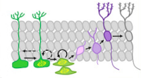
Neuronal Exosomes as a New Signaling System
Number of studies devoted to investigation of neuronal exosomes increases significantly each year. Potential of exosomes as diagnostic markers of neurodegenerative diseases has been examined thoroughly and similar protocols were used to search for the markers of other psychiatric disorders. Biogenesis of exosomes in various types of cells has been studied, physiological role of exosomes has been actively investigated, and many features of their signaling cascades have been clarified. The accumulated data indicate important role of the exosome signaling in interneuronal communication. Do we have enough grounds to recognize exosomes as new non-canonical neurotransmitters in the brain? In this review we discuss this issue and present a concept on the possible role of brain exosomes as a new signaling system to the scientific community.
-
Delayed TBI-Induced Neuronal Death in the Ipsilateral Hippocampus and Behavioral Deficits in Rats: Influence of Corticosterone-Dependent Survivorship Bias?
Acute and chronic corticosterone (CS) elevations after traumatic brain injury (TBI) may be involved in distant hippocampal damage and the development of late posttraumatic behavioral pathology. CS-dependent behavioral and morphological changes were studied 3 months after TBI induced by lateral fluid percussion in 51 male Sprague–Dawley rats. CS was measured in the background 3 and 7 days and 1, 2 and 3 months after TBI. Tests including open field, elevated plus maze, object location, new object recognition tests (NORT) and Barnes maze with reversal learning were used to assess behavioral changes in acute and late TBI periods. The elevation of CS on day 3 after TBI was accompanied by early CS-dependent objective memory impairments detected in NORT. Blood CS levels > 860 nmol/L predicted delayed mortality with an accuracy of 0.947. Ipsilateral neuronal loss in the hippocampal dentate gyrus, microgliosis in the contralateral dentate gyrus and bilateral thinning of hippocampal cell layers as well as delayed spatial memory deficits in the Barnes maze were revealed 3 months after TBI. Because only animals with moderate but not severe posttraumatic CS elevation survived, we suggest that moderate late posttraumatic morphological and behavioral deficits may be at least partially masked by CS-dependent survivorship bias.
-
A Comparative Study of Two Models of Intraluminal Filament Middle Cerebral Artery Occlusion in Rats: Long-Lasting Accumulation of Corticosterone and Interleukins in the Hippocampus and Frontal Cortex in Koizumi Model
Recently, we have shown the differences in the early response of corticosterone and inflammatory cytokines in the hippocampus and frontal cortex (FC) of rats with middle cerebral artery occlusion (MCAO), according to the methods of Longa et al. (LM) and Koizumi et al. (KM) which were used as alternatives in preclinical studies to induce stroke in rodents. In the present study, corticosterone and proinflammatory cytokines were assessed 3 months after MCAO. The most relevant changes detected during the first days after MCAO became even more obvious after 3 months. In particular, the MCAO-KM (but not the MCAO-LM) group showed significant accumulation of corticosterone and IL1β in both the ipsilateral and contralateral hippocampus and FC. An accumulation of TNFα was detected in the ipsilateral hippocampus and FC in the MCAO-KM group. Thus, unlike the MCAO-LM, the MCAO-KM may predispose the hippocampus and FC of rats to long-lasting bilateral corticosterone-dependent distant neuroinflammatory damage. Unexpectedly, only the MCAO-LM rats demonstrated some memory deficit in a one-trial step-through passive avoidance test. The differences between the two MCAO models, particularly associated with the long-lasting increase in glucocorticoid and proinflammatory cytokine accumulation in the limbic structures in the MCAO-KM, should be considered in the planning of preclinical experiments, and the interpretation and translation of received results.
-
Real-time tracking of brain oxygen gradients and blood flow during functional activation
Significance
Cerebral metabolic rate of oxygen (CMRO2) consumption is a key physiological variable that characterizes brain metabolism in a steady state and during functional activation.
Aim
We aim to develop a minimally invasive optical technique for real-time measurement of CMRO2 concurrently with cerebral blood flow (CBF).
Approach
We used a pair of macromolecular phosphorescent probes with nonoverlapping optical spectra, which were localized in the intra- and extravascular compartments of the brain tissue, thus providing a readout of oxygen gradients between these two compartments. In parallel, we measured CBF using laser speckle contrast imaging.
Results
The method enables computation and tracking of CMRO2 during functional activation with high temporal resolution (∼7 Hz). In contrast to other approaches, our assessment of CMRO2 does not require measurements of CBF or hemoglobin oxygen saturation.
Conclusions
The independent records of intravascular and extravascular partial pressures of oxygen, CBF, and CMRO2 provide information about the physiological events that accompany neuronal activation, creating opportunities for dynamic quantification of brain metabolism.
Elevated Serum Cortisol Levels in Patients with Focal Epilepsy, Depression, and Comorbid Epilepsy and Depression
Background: The hypothalamic-pituitary-adrenal (HPA) axis, inflammatory processes and neurotrophic factor systems are involved in pathogenesis of both epilepsy and depressive disorders. The study aimed to explore these systems in patients with focal epilepsy (PWE, n = 76), epilepsy and comorbid depression (PWCED n = 48), and major depressive disorder (PWMDD, n = 62) compared with healthy controls (HC, n = 78). Methods: Parameters of the HPA axis, neurotrophic factors, and TNF-α were measured in blood serum along with the hemogram. Results: Serum cortisol level was augmented in PWE, PWCED, and PWMDD compared with HC and was higher in PWMDD than in PWE. Serum cortisol negatively correlated with Mini–Mental State Examination (MMSE) score in PWE, and positively with depression inventory–II (BDI-II) score in PWMDD. Only PWMDD demonstrated elevated plasma ACTH. Serum TNF-α, lymphocytes, and eosinophils were augmented in PWMDD; monocytes elevated in PWE and PWCED, while neutrophils were reduced in PWE and PWMDD. Serum BDNF was decreased in PWE and PWCED, CNTF was elevated in all groups of patients. In PWE, none of above indices depended on epilepsy etiology. Conclusions: The results confirm the involvement of HPA axis and inflammatory processes in pathogenesis of epilepsy and depression and provide new insights in mechanisms of epilepsy and depression comorbidity.
Detailed Analysis of Dorsal-Ventral Gradients of Gene Expression in the Hippocampus of Adult Rats
We performed RNA sequencing of the dorsal and ventral parts of the hippocampus and compared it with previously published data to determine the differences in the dorsoventral gradients of gene expression that may result from biological or technical variability. Our data suggest that the dorsal and ventral parts of the hippocampus differ in the expression of genes related to signaling pathways mediated by classical neurotransmitters (glutamate, GABA, monoamines, etc.) as well as peptide and Wnt ligands. These hippocampal parts also diverge in the expression of axon-guiding molecules (both receptors and ligands) and splice isoforms of genes associated with intercellular signaling and cell adhesion. Furthermore, analysis of differential expressions of genes specific for astrocytes, microglia, oligodendrocytes, and vascular cells suggests that non-neuronal cells may also differ in the characteristics between hippocampal parts. Analysis of expression of transposable elements showed that depletion of ribosomal RNA strongly increased the representation of transposable elements in the RNA libraries and helped to detect a weak predominance of expression of these elements in the ventral hippocampus. Our data revealed new molecular dimensions of functional differences between the dorsal and ventral hippocampus and points to possible cascades that may be involved in the longitudinal organization of the hippocampus.
Amyloid Aβ25-35 Aggregates Say ‘NO’ to Long-Term Potentiation in the Hippocampus through Activation of Stress-Induced Phosphatase 1 and Mitochondrial Na+/Ca2+ Exchanger
The search for strategies for strengthening the synaptic efficiency in Aβ25-35-treated slices is a challenge for the compensation of amyloidosis-related pathologies. Here, we used the recording of field excitatory postsynaptic potentials (fEPSPs), nitric oxide (NO) imaging, measurements of serine/threonine protein phosphatase (STPP) activity, and the detection of the functional mitochondrial parameters in suspension of brain mitochondria to study the Aβ25-35-associated signaling in the hippocampus. Aβ25-35 aggregates shifted the kinase–phosphatase balance during the long-term potentiation (LTP) induction in the enhancement of STPP activity. The PP1/PP2A inhibitor, okadaic acid, but not the PP2B blocker, cyclosporin A, prevented Aβ25-35-dependent LTP suppression for both simultaneous and delayed enzyme blockade protocols. STPP activity in the Aβ25-35-treated slices was upregulated, which is reverted relative to the control values in the presence of PP1/PP2A but not in the presence of the PP2B blocker. A selective inhibitor of stress-induced PP1α, sephin1, but not of the PP2A blocker, cantharidin, is crucial for Aβ25-35-mediated LTP suppression prevention. A mitochondrial Na+/Ca2+ exchanger (mNCX) blocker, CGP37157, also attenuated the Aβ25-35-induced LTP decline. Aβ25-35 aggregates did not change the mitochondrial transmembrane potential or reactive oxygen species (ROS) production but affected the ion transport and Ca2+-dependent swelling of organelles. The staining of hippocampal slices with NO-sensitive fluorescence dye, DAF-FM, showed stimulation of the NO production in the Aβ25-35-pretreated slices at the dendrite-containing regions of CA1 and CA3, in the dentate gyrus (DG), and in the CA1/DG somata. NO scavenger, PTIO, or nNOS blockade by selective inhibitor 3Br-7NI partly restored the Aβ25-35-induced LTP decline. Thus, hippocampal NO production could be another marker for the impairment of synaptic plasticity in amyloidosis-related states, and kinase–phosphatase balance management could be a promising strategy for the compensation of Aβ25-35-driven deteriorations.
Contribution of histone acetylation to the serotonin-mediated long-term synaptic plasticity in terrestrial snails
Serotonin plays a decisive role in long-term synaptic plasticity and long-term memory in mollusks. Previously, we demonstrated that histone acetylation is a regulatory mechanism of long-term memory in terrestrial snail. At the behavioral level, many studies were done in Helix to elucidate the role of histone acetylation and serotonin. However, the impact of histone acetylation on long-term potentiation of synaptic efficiency in electrophysiological studies in Helix has been studied only in one paper. Here we investigated effects of serotonin, histone deacetylases inhibitors sodium butyrate and trichostatin A, and a serotonergic receptor inhibitor methiothepin on long-term potentiation of synaptic responses in vitro. We demonstrated that methiothepin drastically declined the EPSPs amplitudes when long-term potentiation was induced, while co-application either of histone deacetylase inhibitors sodium butyrate or trichostatin A with methiothepin prevented the weakening of potentiation. We showed that single serotonin application in combination with histone deacetylase blockade could mimic the effect of repeated serotonin applications and be enough for sustained long-lasting synaptic changes. The data obtained demonstrated that histone deacetylases blockade ameliorated deficits in synaptic plasticity induced by different paradigms (methiothepin treatment, the weak training protocol with single application of serotonin), suggesting that histone acetylation contributes to the serotonin-mediated synaptic plasticity.
A Comparative Study of Koizumi and Longa Methods of Intraluminal Filament Middle Cerebral Artery Occlusion in Rats: Early Corticosterone and Inflammatory Response in the Hippocampus and Frontal Cortex
Two classical surgical approaches for intraluminal filament middle cerebral artery occlusion (MCAO), the Longa et al. (LM) and Koizumi et al. methods (KM), are used as alternatives in preclinical studies to induce stroke in rodents. Comparisons of these MCAO models in mice showed critical differences between them along with similarities (Smith et al. 2015; Morris et al. 2016). In this study, a direct comparison of MCAO-KM and MCAO-LM in rats was performed. Three days after MCAO, infarct volume, mortality rate, neurological deficit, and weight loss were similar in these models. MCAO-LM rats showed an increase in ACTH levels, while MCAO-KM rats demonstrated elevated corticosterone and interleukin-1β in blood serum. Corticosterone accumulation was detected in the frontal cortex (FC) and the hippocampus of the MCAO-KM group. IL1β beta increased in the ipsilateral hippocampus in the MCAO-KM group and decreased in the contralateral FC of MCAO-LM rats. Differences revealed between MCAO-KM and MCAO-LM suggest that corticosterone and interleukin-1β release as well as hippocampal accumulation is more expressed in MCAO-KM rats, predisposing them to corticosterone-dependent distant neuroinflammatory hippocampal damage. The differences between two models, particularly, malfunction of the hypothalamic-pituitary-adrenal axis, should be considered in the interpretation, comparison, and translation of pre-clinical experimental results.
Brain Trauma, Glucocorticoids and Neuroinflammation: Dangerous Liaisons for the Hippocampus
Glucocorticoid-dependent mechanisms of inflammation-mediated distant hippocampal damage are discussed with a focus on the consequences of traumatic brain injury. The effects of glucocorticoids on specific neuronal populations in the hippocampus depend on their concentration, duration of exposure and cell type. Previous stress and elevated level of glucocorticoids prior to pro-inflammatory impact, as well as long-term though moderate elevation of glucocorticoids, may inflate pro-inflammatory effects. Glucocorticoid-mediated long-lasting neuronal circuit changes in the hippocampus after brain trauma are involved in late post-traumatic pathology development, such as epilepsy, depression and cognitive impairment. Complex and diverse actions of the hypothalamic–pituitary–adrenal axis on neuroinflammation may be essential for late post-traumatic pathology. These mechanisms are applicable to remote hippocampal damage occurring after other types of focal brain damage (stroke, epilepsy) or central nervous system diseases without obvious focal injury. Thus, the liaisons of excessive glucocorticoids/dysfunctional hypothalamic–pituitary–adrenal axis with neuroinflammation, dangerous to the hippocampus, may be crucial to distant hippocampal damage in many brain diseases. Taking into account that the hippocampus controls both the cognitive functions and the emotional state, further research on potential links between glucocorticoid signaling and inflammatory processes in the brain and respective mechanisms is vital.
The Role of Transposable Elements of the Human Genome in Neuronal Function and Pathology
Transposable elements (TEs) have been extensively studied for decades. In recent years, the introduction of whole-genome and whole-transcriptome approaches, as well as single-cell resolution techniques, provided a breakthrough that uncovered TE involvement in host gene expression regulation underlying multiple normal and pathological processes. Of particular interest is increased TE activity in neuronal tissue, and specifically in the hippocampus, that was repeatedly demonstrated in multiple experiments. On the other hand, numerous neuropathologies are associated with TE dysregulation. Here, we provide a comprehensive review of literature about the role of TEs in neurons published over the last three decades. The first chapter of the present review describes known mechanisms of TE interaction with host genomes in general, with the focus on mammalian and human TEs; the second chapter provides examples of TE exaptation in normal neuronal tissue, including TE involvement in neuronal differentiation and plasticity; and the last chapter lists TE-related neuropathologies. We sought to provide specific molecular mechanisms of TE involvement in neuron-specific processes whenever possible; however, in many cases, only phenomenological reports were available. This underscores the importance of further studies in this area.A Combined Effect of G-Quadruplex and Neuro-Inducers as an Alternative Approach to Human Glioblastoma Therapy
Cancer cell reprogramming based on treatment with G-quadruplex, having antiproliferative power, along with small molecules able to develop iPSCs into neurons, could create a novel approach to diminish the chance of glioblastoma recurrence and circumvent tumor resistance to conventional therapy. In this research, we have tested several combinations of factors to affect both total cell cultures, derived from tumor tissue of patients after surgical resection and two subfractions of this cell culture after dividing them into CD133-enriched and CD133-depleted populations (assuming CD133 to be a marker of glioblastoma stem-like cells). CD133+ and CD133- cells exhibit different responses to the same combinations of factors; CD133+ cells have stem-like properties and are more resistant. Therefore, the ability to affect CD133+ cells provides a possibility to circumvent resistance to conventional therapy and to build a promising strategy for translation to improve the treatment of patients with glioblastoma.
Neuroinflammation and Neuronal Loss in the Hippocampus Are Associated with Immediate Posttraumatic Seizures and Corticosterone Elevation in Rats
Hippocampal damage after traumatic brain injury (TBI) is associated with late posttraumatic conditions, such as depression, cognitive decline and epilepsy. Mechanisms of selective hippocampal damage after TBI are not well understood. In this study, using rat TBI model (lateral fluid percussion cortical injury), we assessed potential association of immediate posttraumatic seizures and changes in corticosterone (CS) levels with neuroinflammation and neuronal cell loss in the hippocampus. Indices of distant hippocampal damage (neurodegeneration and neuroinflammation) were assessed using histological analysis (Nissl staining, Iba-1 immunohistochemical staining) and ELISA (IL-1β and CS) 1, 3, 7 and 14 days after TBI or sham operation in male Wistar rats (n = 146). IL-1β was elevated only in the ipsilateral hippocampus on day 1 after trauma. CS peak was detected on day 3 in blood, the ipsilateral and contralateral hippocampus. Neuronal cell loss in the hippocampus was demonstrated bilaterally; in the ipsilateral hippocampus it started earlier than in the contralateral. Microglial activation was evident in the hippocampus bilaterally on day 7 after TBI. The duration of immediate seizures correlated with CS elevation, levels of IL-1β and neuronal loss in the hippocampus. The data suggest potential association of immediate post-traumatic seizures with CS-dependent neuroinflammation-mediated distant hippocampal damage.
Potassium channels as prominent targets and tools for the treatment of epilepsy
K+ channels are of great interest to epilepsy research as mutations in their genes are found in humans with inherited epilepsy. At the level of cellular physiology, K+ channels control neuronal intrinsic excitability and are the main contributors to membrane repolarization of active neurons. Recently, a genetically modified voltage-dependent K+ channel has been patented as a remedy for epileptic seizures.
Neural versus alternative integrative systems: molecular insights into origins of neurotransmitters
Transmitter signalling is the universal chemical language of any nervous system, but little is known about its early evolution. Here, we summarize data about the distribution and functions of neurotransmitter systems in basal metazoans as well as outline hypotheses of their origins. We explore the scenario that neurons arose from genetically different populations of secretory cells capable of volume chemical transmission and integration of behaviours without canonical synapses. The closest representation of this primordial organization is currently found in Placozoa, disk-like animals with the simplest known cell composition but complex behaviours. We propose that injury-related signalling was the evolutionary predecessor for integrative functions of early transmitters such as nitric oxide, ATP, protons, glutamate and small peptides. By contrast, acetylcholine, dopamine, noradrenaline, octopamine, serotonin and histamine were recruited as canonical neurotransmitters relatively later in animal evolution, only in bilaterians. Ligand-gated ion channels often preceded the establishment of novel neurotransmitter systems. Moreover, lineage-specific diversification of neurotransmitter receptors occurred in parallel within Cnidaria and several bilaterian lineages, including acoels. In summary, ancestral diversification of secretory signal molecules provides unique chemical microenvironments for behaviour-driven innovations that pave the way to complex brain functions and elementary cognition.
Glucocorticoid‐mediated mechanisms of hippocampal damage: Contribution of subgranular neurogenesis
Our employees from the Laboratory of Functional Biochemistry of Nervous System presented a comprehensive overview of the interplay between glucocorticoids (GCs) and adult hippocampal neurogenesis (AHN), particularly, in the context of a diseased brain. The effectors of GCs in the dentate gyrus neurogenic niche of the hippocampal are reviewed, and the consequences of the GC signaling on the generation and integration of new neurons are discussed. Recent findings demonstrating how GC signaling mediates impairments of the AHN in various brain pathologies are overviewed. GC‐mediated effects on the generation and integration of adult‐born neurons in the hippocampal dentate gyrus depend on the nature, severity, and duration of the acting stress factor. GCs realize their effects on the AHN primarily via specific glucocorticoid and mineralocorticoid receptors. Disruption of the reciprocal regulation between the hypothalamic–pituitary–adrenal (HPA) axis and the generation of the adult‐born granular neurons is currently considered to be a key mechanism implicating the AHN into the pathogenesis of numerous brain diseases, including those without a direct hippocampal damage. These alterations vary from reduced proliferation of stem and progenitor cells to increased cell death and abnormalities in morphology, connectivity, and localization of young neurons. Although the involvement of the mutual regulation between the HPA axis and the AHN in the pathogenesis of cognitive deficits and mood impairments is evident, several unresolved critical issues are stated. Understanding the details of GC‐mediated mechanisms involved in the alterations in AHN could enable the identification of molecular targets for ameliorating pathology‐induced imbalance in the HPA axis/AHN mutual regulation to conquer cognitive and psychiatric disturbances.